Around 80,000 cruciate ligaments tear in Germany every year. That's a cruciate ligament tear every 6.5 minutes. This makes the cruciate ligament the most well-known ligament of the knee joint. Since the knee joint is exposed to enormous loads in sports, it is one of the two most injury-prone joints along with the shoulder joint. The knee can be injured through opponent contact in sports such as soccer, ice hockey, and handball, or without the impact of an opponent such as skiing. It is estimated that well over a million cruciate ligament ruptures per year worldwide.
What is the cruciate ligament?
Each knee has two cruciate ligaments, one posterior and one anterior. In principle, both ligaments can tear, but injuries to the anterior cruciate ligament are much more common. Sports accidents are the most common cause here. It is not uncommon for neighboring structures in the knee joint, such as the collateral ligaments or the menisci, to be affected.
The two cruciate ligaments cross in the center of the knee joint, hence their name. The primary role is secondarily to stabilize the knee joint in rotation and ground contact in all sports involving running, sprinting and jumping. They are particularly stressed during quick changes of direction, in which the standing leg is fixed and the upper body is already moving in a different direction.
The cruciate ligaments are fibrous structures that require considerable forces to tear. The cruciate ligament has a tear strength of around 2400 kg. However, the tear strength of the cruciate ligament varies: the diameter is smaller in women in particular, which is one of the main reasons why they are more frequently affected by cruciate ligament tears. Studies have shown that the risk in women is two to eight times greater than in men. This has to do with the already mentioned relationship between the front and back of the thigh, but there are also hormonal causes and also anatomical reasons.
The thicker the cruciate ligament, the better
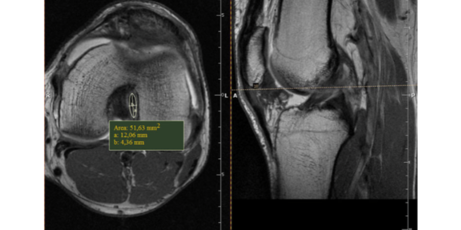
A primary anatomical reason for cruciate ligament tears is the thickness of the cruciate ligament. A study (1) by Grzelak et al from 2012 gives a very interesting perspective on the topic of hypertrophy ( engl. thickening ) of the cruciate ligaments. This came to the conclusion that strength training increases the thickness of the cruciate ligaments by up to 350%. In this study, the thickness of the two cruciate ligaments was compared to that of a control group based on MRI images of weightlifters. The result was a significant difference. The study was the first scientific study to demonstrate cruciate ligament hypertrophy. It was also found that the earlier strength training was started, ideally before puberty, the greater the effect of strengthening the cruciate ligaments. An interesting hypothesis of the study regarding the development of the cruciate ligaments is that the artery that supplies the cruciate ligaments (arteria genus media or "middle knee artery" ) recedes in the course of adolescence and thus the supply of nutrients to the cruciate ligaments in children and adolescents is much better guaranteed.
The results of the study (1) here:
The study also states that one of the reasons for this hypertrophy of the cruciate ligaments is that when lifting weights, the knee is subjected to high resistance beyond maximum flexion. This maximum flexion under high forces and the associated stretching of the cruciate ligaments activates the fibroblasts, which initiate ligament hypertrophy.
The study clearly shows that strength training with high resistance results not only in hypertrophy of the muscles but also in hypertrophy of the cruciate ligaments. And thus offers an interesting perspective on the pre- and rehabilitation of cruciate ligament tears.
Good luck with strength training and cruciate ligament hypertrophy!
Reference:
(1) International Orthopedics (SICOT) (2012) 36:1715-1719; Hypertrophied cruciate ligament in high performance weightlifters observed in magnetic resonance imaging; Piotr Grzelak & Michał Podgorski & Ludomir Stefanczyk & Marek Krochmalski & Marcin Domzalski; Received: 17 January 2012 / Accepted: 4 March 2012 / Published online: 25 March 2012 # The Author(s) 2012. This article is published with open access at Springerlink.com https://www.ncbi.nlm.nih.gov /pmc/articles/PMC3535026/
Image: MRI images of the cruciate ligaments (Source: see reference)


